Menu
Log in


Log in

Login

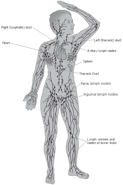
WHAT IS LYMPHEDEMA?

Lymphedema is chronic swelling (edema) caused by a buildup of fluid (lymph).

This happens when the lymphatic system is either faulty or damaged and cannot function as normal. It leads to chronic (long-lasting) swelling in the tissues where the lymph flow is blocked. Lymphedema most commonly presents in a limb  (arm/hand or leg/foot), but it can also be present in the breast, trunk, genitals, or head and neck.


> Lymphatic System

> Signs & Symptoms

> Stages

> Diagnosis

> Diagnostic Tests

> Certified Lymphedema Therapists (Treatment options)

> Surgical Options

> Risk reduction strategies

> Infections


LYMPHATIC SYSTEM


The lymphatic system is an important part of the immune system.

USEFUL LINKS TO LEARN MORE ABOUT THE LYMPHATIC SYSTEM

The Lymphatic System

https://www.cdc.gov/cancer/survivors/patients/lymphedema.htm

Lymph Vessel System

http://www.vodderschool.com/lymph_vessel_system


It is estimated that 1 million Canadians have the incurable disease, many unaware of the true cause of their swelling which is often misinterpreted as fat. There are many causes of lymphedema classified into two forms:

Primary lymphedema is caused by a defect of the lymphatic system.  It may be present at birth, develop when puberty begins or in adulthood.


Secondary lymphedema is caused due to an event that damages or blocks part of the lymphatic system. The most common cause is the result of treatment for some cancers (surgery, radiation).  Other causes of damage to the lymphatic system include trauma caused by an accident, injury or a burn.


SIGNS AND SYMPTOMS


·       Abnormal swelling in the affected body part

·       Full or heavy feeling in the affected body part

·       Decreased mobility or achy in the affected body part

·       Clothing, jewellery or shoes may feel tighter but not due to weight gain


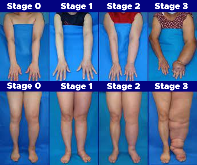
If you feel unwell or notice a sign of infection such as increased redness, swelling, warmth or pain to the affected limb, you should be seen by your doctor or emergency personnel immediately.  At present there is no cure for lymphedema; however, it can be successfully managed if diagnosed and treated in a timely man


STAGES

Stages help classify the severity of Lymphedema.  Currently there are four (4) known stages ranging from mild to severe. 

Different methods are used worldwide to classify the stages.  However, you can perform a simple pitting edema test to give you a rough idea (relevant for Stage 1).

Stage 0 (latent) – The least severe of all and not truly visible.

Stage 1 (early or mild) – Lymphedema is reversible as the swelling will disappear with elevation or bedrest.

Stage 2 (moderate) – Elevating does not resolve swelling. 
May or may not be pitting.  Fat accumulation of fibrosis starts.

Stage 3 (severe) – Most advanced stage. 
Fibrosis is hard and difficult to soften. The limb(s) can be misshapen or large.
 You are more susceptible to infections and wounds.


LYMPHEDEMA DIAGNOSIS

Your family doctor may not be able to diagnose Lymphedema (yet).  Training is available for doctors if they wish to know more about this disease.

For doctor training:

https://www.ualberta.ca/rehabilitation/programs/professional-development/microcredential-courses/lymphedema-and-chronic-edema.html


DOCTORS FOR LYMPHEDEMA DIAGNOSIS & TREATMENT


DR ELLIOTT WEISS, MD FRCPC

Consultant Physical Medicine & Rehabilitation Providence Health Care 408-1160 Burrard St Vancouver BC V6Z 1Y6

Fax Referral to: 604-974-8882 | Email: eweiss@providencehealth.bc.ca


LAWRENCE KEI, MD. MLD 

Opal Physiotherapy Clinic

20226 Fraser Hwy #102, Langley BC V3A4E6

Fax Referral to: 604-532-7897


DR ERIN BROWN, MD, PhD, FRCSC

UBC/VGH Lymphedema Program - Division Head

Gordon & Leslie Diamond Health Care Centre Plastic Surgery, 3rd floor, Reception 8

2775 Laurel Street, Vancouver BC V5Z 1M9 Tel: 604.875.5866 ext 2

Fax Referral to: 604-875.5861

DIAGNOSTIC TESTS

The Stemmer sign is a physical examination. If the examiner cannot pinch the skin of the dorsum of the foot or hand, then this positive finding is associated with lymphedema.

    • Measuring the volume (using a perometer) or circumference (using a measuring tape) of a limb and comparing to the opposite unaffected limb is a physical examination used by a therapist.  
    • A volume of difference of 100 ml between limbs or a 2 cm difference (measured at set intervals along the limb) is often used for a definitive diagnosis of lymphedema.

    • Lymphoscintigraphy is performed by a doctor.  During this test, the person is injected with a radioactive dye and then scanned by a machine.  The resulting images show the dye moving through the lymph vessels, highlighting blockages to confirm Lymphedema.
    • Indocyanine green lymphography (ICG) is performed by a doctor.  During this test, the person is injected with a green colored medical dye and then scanned by a machine.  The resulting image scans the superficial lymphatics and helps delineate individualized lymphedema treatment.

    •  Magnetic Resonance Lymphangiography (MRL) (may not be available in B.C. yet). 

    CERTIFIED LYMPHEDEMA THERAPISTS (TREATMENT OPTIONS):

    Certified Lymphedema therapists are massage therapists, physiotherapists, kinesiologists or nurses who have additional education and training in Complete Decongestive Therapy (CDT) to reduce your swelling, improve the condition of your skin and increase your mobility.  The two phases of CDT are:

    Phase 1: Decongestion which includes:

    Compression Therapy to reduce

    Skin care/wound care

    Manual Lymphatic Drainage (MLD)

    Self-Management

    Exercise movement


    Phase 2: Maintenance which includes:

    Compression Therapy to maintain

    Skin care/wound care

    Manual Lymphatic Drainage (MLD)

    Self-Management

    Exercise movement


    The therapist will educate you on lymphedema management best practices you can do on your own including: Manual Lymph Drainage, Compression, Skin Care and Exercise.  A therapist may also recommend you to a certified garment fitter for compression.


    Online Professional Listing

    SURGICAL OPTIONS

    As of 2023, there is currently no cure for Lymphedema, but research into the lymphatic system is progressing around the world.  Surgical options may help improve your daily living if you have been managing your lymphedema with Complete Decongestive Treatment with no improvement.  

    The UBC/VGH Lymphedema clinic doctors will provide a comprehensive assessment to provide the most accurate diagnosis and staging, to maximize conservative treatment and to determine if surgical treatment may be beneficial. 

    The UBC/VGH Lymphedema clinic provides the following types of surgeries to B.C. patients:

    Lymphaticovenous Anastomosis (LVA) is a method of redirecting excess lymphatic fluid into the venous system to reduce the appearance of lymphedema and prevent its recurrence.  This is the least invasive surgery for lymphedema. LVA is microsurgery too.

    Vascularized Lymph Node Transfer (VLNT) is a microsurgical procedure where lymph nodes are transferred from a low risk lymph node basin to a limb with symptomatic lymphedema.

    Liposuction or Suction-assisted Protein Lipectomy (SAPL) should only be performed by surgeons trained to treat lymphedema to not further damage the lymphatic system.  This procedure is done under general anaesthetic to suction out the fat caused by Lymphedema.  Patients still need to continue with ongoing lymphedema management best practices (including wearing compression garments) after this surgery.

     
    WHO IS AT RISK FOR LYMPHEDEMA 

    Your risk for lymphedema depends on a number of factors, including:

    ● A family history of chronic (long-lasting) swelling

    ● If you had surgery, the number, size and location of lymph nodes removed or damaged

    ● If you had radiation therapy or an injury, the degree of damage to your lymphatic system

    ● Having chronic venous disease

    ● Having a history of skin infections, including cellulitis

    ● Being overweight and/or inactive add to these risk factors


    Having a risk factor doesn’t mean you’re going to develop lymphedema but if your lymphatic system is faulty or damaged, you have a lifetime risk for lymphedema.

    We can’t predict who’s going to develop lymphedema. Your body may heal well after treatment and be able to manage the lymph flow in the damaged area. Or, the extent of damage to your lymphatic system may combine with other risk factors and trigger lymphedema during treatment or many years later.

    Risk Reduction strategies when living with lymphedema:

    • Practice good skin care

    Keep your skin clean, soft and moisturized to avoid skin infections.

    Take care of scratches, pinpricks, cuts, burns or splinters right away.

    Get treated for skin problems, including inflammation, fungal infections, ingrown toenails or dermatitis.

    Avoid skin punctures, blood tests or vaccination shots on side where your lymphatic system may be damaged.

    • Exercise regularly at a moderate pace
    • Avoid sitting or standing for long periods
    • Long periods of travel may require compression garments
    • Aim for a healthy body weight
    • Avoid high temperatures

    Stay indoors (ideally with air conditioning) in the high heat and humidity of summer, stay away from hot tubs/saunas and take warm, rather than hot, baths and showers.

    • Avoid tight clothing or jewelry

    You may find it helpful to speak to a certified lymphedema therapist about your risk for lymphedema and ways to lower it.

    INFECTIONS


    Introduction

    Serious infections that can develop within the affected tissues are a serious complication associated with lymphedema.  The risk of infection increases when lymphedema is not controlled by conservative management strategies and appropriate precautions to prevent infection entering through any wound or puncture.


    The risks of lymphedema related infections are due to:

    • The swelling of lymphedema compromises the health of the skin.  Healthy intact skin is the body’s primary line of defense against invading pathogens.  Normal skin is protected by a film known as the acid mantle.  The acidic nature of this film discourages such pathogens.  When skin is swollen, the acid mantle is disrupted and is not as effective in stopping invading pathogens.
    • Protein-rich, stagnant lymph within these swollen tissues creates an environment that pathogens love!  This lymph has nutrients that allow the pathogens to thrive.  This stagnant lymph can also contain pathogens and damaging toxins that would normally hae been removed with  the normal flow of lymph.
    • The deep skin folds resulting from the lymphedema are an ideal breeding ground for fungal infections.  The area within the folds are warm, moist, and dark.  This creates an ideal environment for fungi such as tinea pedis (athlete's foot) and tinea cruris (jock itch).

    Erysipelas 

    Erysipelas is an acute infection typically with a skin rash, usually on any of the legs and toes, face, arms, and fingers. It is an infection of the upper dermis and superficial lymphatics, usually caused by A Streptococcus bacteria on scratches or otherwise infected areas. Erysipelas is more superficial than

    cellulitis, and is typically more raised and demarcated. Signs and symptoms may include high fever, chills, shaking, headaches, fatigue or vomiting; a general feeling of un-wellness. The rash is typically red, warm and can be painful. It can change and grow in surface area in a very short period of time (a few hours to 48 hours)

    Cellulitis 
    Cellulitis is a bacterial infection involving the inner layers of the skin. It specifically affects the dermis and subcutaneous fat. Signs and symptoms include an area of redness which increases in size over a few days. The area of redness may be blotchy with irregular borders, there may be swelling in the area.
     While the redness often turns white when pressure is applied, this is not always the case. The area of infection is usually painful, and the person may have a fever and feel tired. It is an aggressive infection which can affect lymphatic transport capacity. Group A Streptococcus and Staphylococcus are the most common of these bacteria, which are part of the normal flora of the skin, which normally cause no actual infection while on the skin's outer surface. If you are suspicious about the possibility of either of these infections, please see your doctor immediately. A course of antibiotics either oral or via injection is necessary to control the infection. 

    Printable Resources for Cellulitis
    Cellulitis & Lymphedema ~ What You Need to Know
    Lymphedema and the Risk of Infection

      Lymphangitis

      Lymphangitis (lim-fan-JIGH-tis) is an infection involving the lymphatic vessels that is most commonly caused by the spreading of an acute streptococcal or staphylococcal infection of the skin.  The presence of lymphangitis suggests that an infection is progressing and should raise concerns of spread of bacteria to the bloodstream.


      Known as sepsis, a bacterial infection in the bloodstream can spread to all of the body systems within a matter of hours.  Therefore, at the first signs of lymphangitis, you should seek medical treatment immediately.


      Symptoms of Lymphangitis:

      • Malaise, loss of appetite, headache, and muscle aches
      • Red streaks from infected area to the armpit or groin (these may be faint or obvious)
      • Swollen lymph nodes
      • Chills and fever

      Sweets Syndrome
      Sweet's syndrome undefined also known as acute febrile neutrophilic dermatosis undefined is a rare skin condition marked by fever and painful skin lesions (raised papules that appear mainly on your arms, neck, face and back.

      The exact cause of Sweet's syndrome isn't always known. In some people, it's triggered by an infection, illness or certain medications. Sweet's syndrome can also occur with some types of cancer.

      The most common treatment for Sweet's syndrome is corticosteroid pills, such as prednisone. Signs and symptoms often disappear just a few days after treatment begins, but recurrence is common.

      Sweet's syndrome is marked by an abrupt eruption of small red bumps on your arms, neck, face or back — often after a fever or upper respiratory infection. The bumps grow quickly in size, spreading into painful clusters up to an inch or so in diameter and treatment should be sought promptly.

      Fungal Infections

      Fungal infections occur most often when the genitalia, legs and feet are affected by stage 2 or stage 3 lymphedema.


      Athlete’s Foot, which is caused by the fungus tinea pedis, occurs on the feet and between the toes.  Jock itch, which is caused by the fungus tinea cruris, thrives in the genital area.  These infections occur when the right combination of conditions exists including:

      • A warm, dark humid environment, such as between the toes.
      • Any change in the health of the skin.
      • Lowering of the body's natural resistance.

      Tinea Pedis Symptoms

      • Pain, burning, and itching
      • Drying, cracking, and scaling of the skin
      • Blistering
      • Swelling
      These infections are difficult to treat and prevention is the best approach, which includes:·  maintain cleanliness by changing shoes and socks as often as necessary controlling moisture by using an antiperspirant powder or spray; routinely using an anti-fungal ointment and/or powder, as recommended by your healthcare provider.

        Jock itch can be treated with over-the-counter ointments; however, it is advisable to see your physician for professional advice.  Once the condition is under control, anti-fungal powders or sprays may be recommended for daily use as a preventive measure.


        References

        Living Well with Lymphedema by A. Ehrlich, A. Vinjé-Harrewijn PT, CLT-LANA, and E. McMahon PhD. Lymph Notes, 2005.

        Lymphedema Diagnosis and Therapy 2-E edited by H. Weissleder and C. Schuehhardt. Kagerer Kommunikation, 1997.

        Lymphedema Management: The comprehensive Guide for Practitioners by J.E. Zuther. Thieme, 2005, pages 249-250.

        The Lymphatic System Pathology by B. Lasinski in Implications for the Physical Therapists 2-Ed by C.C Goodman, W.G. Boissonnault, and K.S.Fuller. Saunders, 2003, pages 477-508.

        Used with permission - © LymphNotes 2008.

        http://www.mayoclinic.org


        This information does not replace the advice of a qualified health care professional.


        Email Address:
        info@bclymph.org
        Telephone:1-604-924-6282 Lower Mainland
        1-866-991-2252 Toll Free
        (Canada & USA)

        Mailing Address Only:

        BC Lymphedema Association
        723 Donegal Place
        North Vancouver, BC  V7N 2X6
        (this is not a lymphedema clinic)

        © 2021 BC Lymphedema Association. | DISCLAIMER | Privacy Policy

        BCLA Logo Designed by RA Designs

        Powered by Wild Apricot Membership Software